
Evaluation of Thoracic and Lumbar Spinal Column Injury. Evaluation and management of vertebral compression fractures. Accessed: March 9, 2017.Īlexandru D, So W. Spinal Column Injuries in Adults: Definitions, Mechanisms, and Radiographs. Trauma of the spine and spinal cord: imaging strategies. Parizel PM, Van der zijden T, Gaudino S, et al. To minimize the risk of spinal cord lesions causing permanent neurological injury, treatment of unstable fractures should be initiated as soon as possible. Kyphoplasty : reexpansion of the fracture through the insertion of an inflatable balloon into the vertebral body and injection of bone cement.Vertebroplasty : injection of bone cement into the fractured vertebra for immediate stabilization.Indication: stable vertebral compression fractures with progressive pain or kyphosis despite conservative treatment.Approach: fusion of two or more vertebral bodies via internal fixation using plates, rods, screws, or cages.Indications: unstable fractures and/or neurological symptoms.

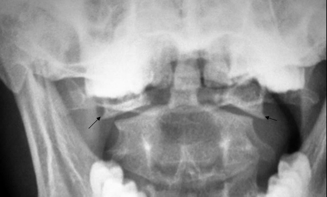
External bracing and orthotics to maintain spinal alignment, promote healing, and control pain through immobilization for about 8–12 weeks (e.g., rigid collar in cervical fracture, cervical-thoracic brace for thoracic fractures, and thoracolumbar-sacral orthosis for lower back fractures).Orotracheal intubation with rapid-sequence intubation is preferred for establishing an airway in an apneic patient with a cervical spine injury.For possible injury of the cervical spine: immobilization with a rigid cervical collar.Place the patient on a long backboard move to stretcher once in the hospital.Rescue from the field when there is concern for vertebral fractures.Treatment: immobilization for stable fractures, surgery for dislocations.Diagnostics: x-ray of the spinal cord to discern an atlantoaxial dislocation, CT, or MRI.Etiology: trauma with hyperextension and distraction (e.g., car accident).Definition: bilateral fracture of the axis arch.Neurological problems ranging from local sensory loss to paralysis due to complete spinal cord injury.A contributing factor is loss of bone substance as a result of osteoporosis (mostly seen in elderly patients).Head or neck injury as a result of a fall or blunt trauma.Epidemiology: 10–15% of all cervical fractures.Definition: fracture of the dens axis (second cervical vertebral body).Treatment: immobilization for stable fractures surgery for dislocations.Arteriography: in cases of vascular compromise.Cervical spine x-ray: fractures and dislocations.An asymptomatic course is also possible.Neurologic deficits, such as Horner syndrome.Neck ache, paravertebral hematoma with dysphagia.Jefferson fracture: combined fracture of the anterior and posterior arches.

Injury mode: axial force (e.g., swimming accident caused by jumping head-first into shallow water).Definition: fracture of the atlas (first cervical vertebra).Fracture- dislocation: fractured vertebra and disrupted ligaments instability may cause spinal cord compression.Possible displacement of bone fragments into the spinal canal.Result of compression trauma with severe axial loading.Burst fracture: fracture of the vertebra in multiple locations.Codfish vertebra: characterized by loss of height of the central part of the vertebral body, resulting in a biconcave vertebral body.Vertebra plana: advanced compression fracture with a loss of height of the entire vertebral body, both anteriorly and posteriorly.Wedge fracture: characterized by a loss of height, predominantly of the anterior part of the vertebral body, which results in a wedge-shaped vertebra.Decreased height (loss of 2–3 cm with each fracture ).Progressive thoracic kyphoticdeformity if multiple vertebrae are affected (stooped posture with a “dowager's hump”).Long-term findings after multiple vertebral compression fractures.


 0 kommentar(er)
